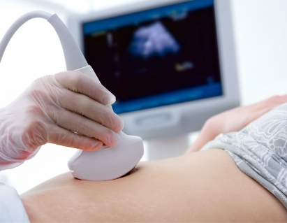
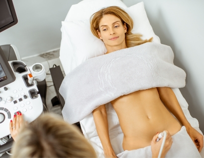
Ultrasound imaging, also known as sonography, is a non-invasive structural screening that uses sound waves instead of radiation to capture live images from inside your body as a proactive way to gain insight into your health.
Services
Ultrasound
• No Pain
• No Radiation
• No Doctor Referral Needed
• Radiologist Read
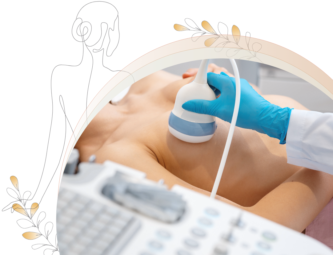
Safe, accurate testing for everyone.

Increased Detection
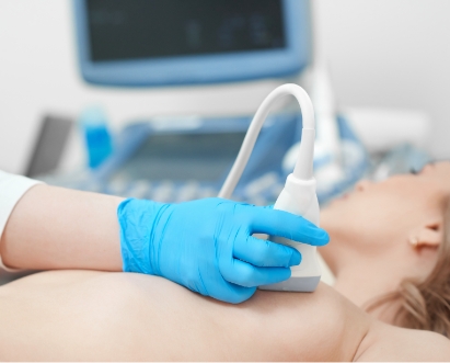
Ultrasound is commonly used to follow up on other abnormal breast screening results. It is an excellent way to determine whether a suspicious lump is a fluid-filled cyst or a solid mass. In women with dense breasts, ultrasound can increase detection from approximately 48% to 97% compared to a mammogram alone since cancer and dense tissue both appear white on a mammogram.

Accessible & Affordable
You do not need a doctor’s referral* or a previous mammogram to schedule an appointment with us. You can schedule screenings with us as often as you wish, at an affordable price. No jumping through hoops with insurance or other physicians. Your health, your way.
*A current diagnosis may require a doctor's order. Please contact us for details.

Reliable & Respected
Ultrasound is a widely accepted and respected form of testing recommended by a variety of practitioners. It is an easy, non-invasive test that can identify areas of concern and can be used as a preventative screening to provide peace of mind. Each breast test has its own strengths and not one is 100% accurate, so it’s important to consider a multimodal approach by utilizing more than one breast screening.
Focus on areas of interest.
Increase your chances of early detection with a widely respected, non-invasive technology.
Breasts & Underarms
Breast ultrasound can help identify suspicious masses in the breast tissue and lymph glands to determine whether a lump is a fluid-filled cyst or a solid mass. This aids in the early discovery of breast cancer and other breast-related conditions like unusual nipple discharge, mastitis, breast pain, redness, and swelling.
Breast ultrasound is highly beneficial for women with dense breast tissue because there are more ducts and glands, more fibrous tissue, and less fatty tissue. Traditional testing is less sensitive to these characteristics. For example, mammograms may not be able to differentiate between breast cancer and dense tissue since they both may appear white on the image. It is estimated that approximately 50% of women have dense breasts and studies have shown that ultrasound significantly increases detection from 48% to 97% compared to mammograms alone.
This may be valuable for:
• Dense breasts
• Implants
• Lumps
• Breast pain
• Pregnant or nursing women
Ultrasound Resources
Breast ultrasound scan

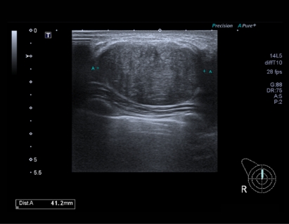
Ultrasound image measuring an invasive breast cancer tumor
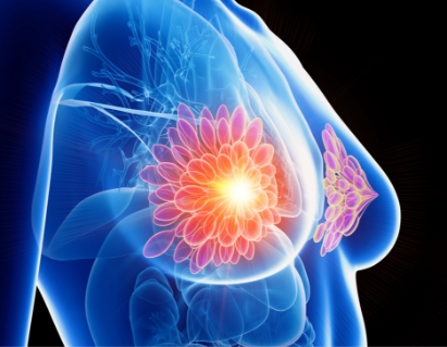
Illustration of breast anatomy
Women’s Health Check
This screening is focused specifically on a woman’s health concerns and needs.
The Women’s Health Check (WHC) includes breasts and pelvic organs and you can choose between the right upper abdomen (liver, pancreas, gallbladder, and right kidney) or thyroid. This can help support your wellness planning and can be used as part of a preventative health program giving you and your practitioner signs of abnormalities before they become serious health issues. The screening may also detect findings that warrant further evaluation and action.
This may be valuable for:
• Breasts
• Pelvic organs
• Abdominal organs
• Thyroid dysfunction
Ultrasound Resources & Articles:

Ultrasound exam
Breast mass
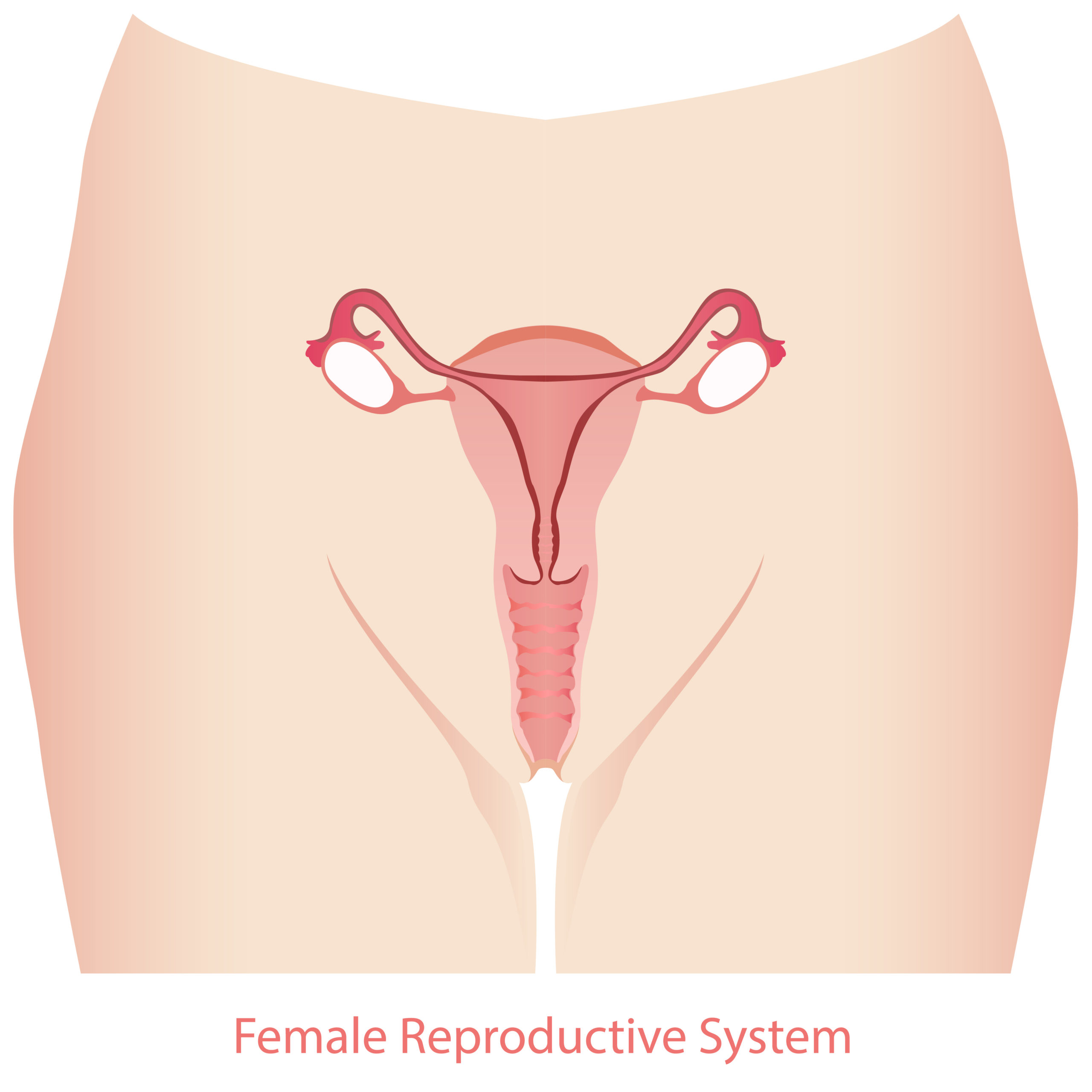
Female reproductive system
Abdomen
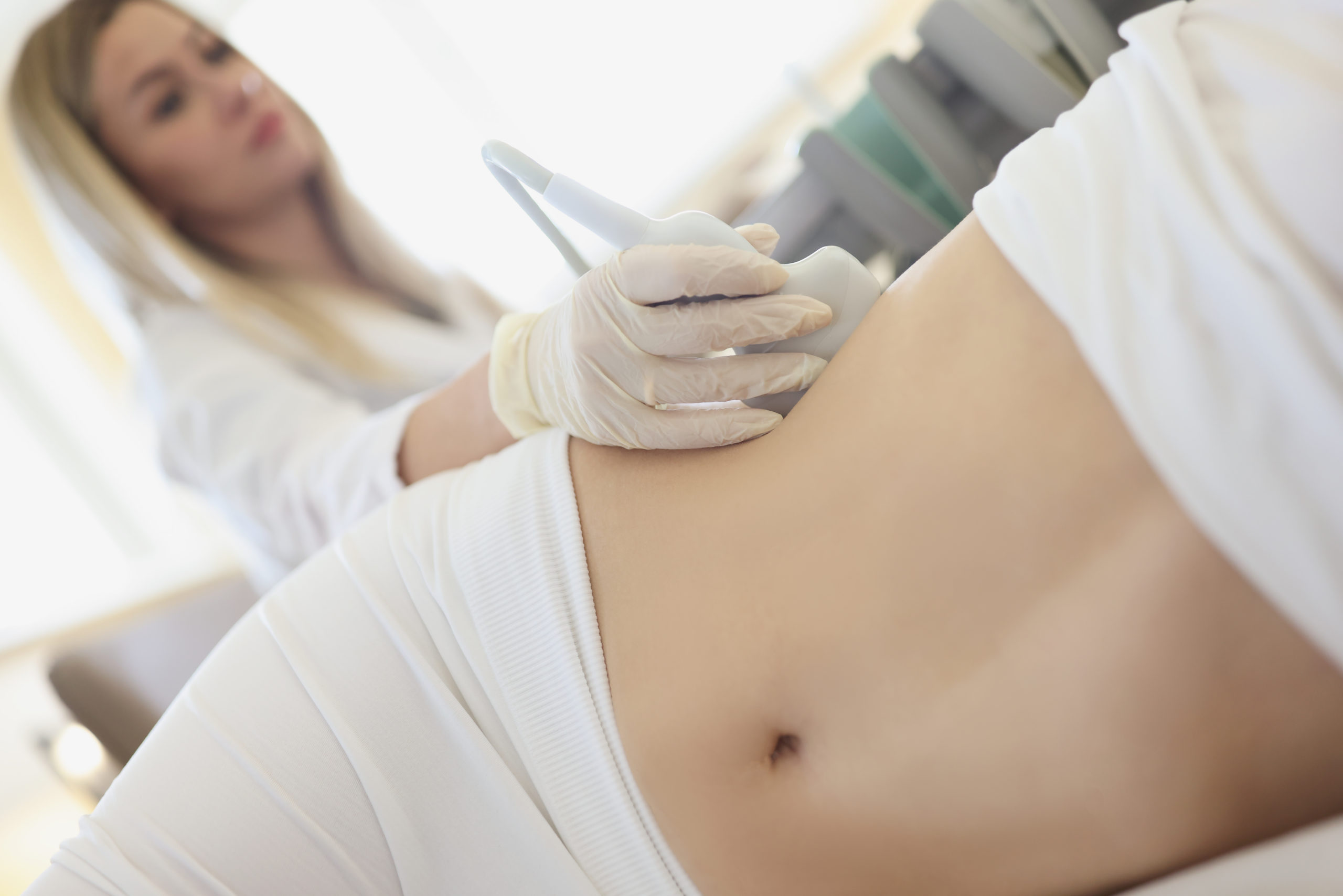
An abdominal ultrasound allows for visualization of the organs and structures. It can help your practitioner evaluate the cause of stomach pain or bloating and help check for kidney stones, liver disease, tumors, and many other conditions. It can also give you peace of mind that your organs are in normal condition and functioning properly. The complete abdominal screening examines the liver, pancreas, gallbladder, spleen, right and left kidney while the right upper abdominal screening examines the liver, pancreas, gallbladder, and right kidney.
The liver supports almost every organ in the body and is vital for survival. It is essential for digesting food and ridding your body of toxic substances. It is also prone to many diseases due to hepatitis, diabetes, fatty liver, cirrhosis from alcohol, and damage from drugs and medications. Symptoms include dark urine, swollen stomach, and excessive fatigue due to loss of nutrients. We screen for structural abnormalities such as abnormal size, cysts, and masses.
The gallbladder is a small pear shaped organ that lies below the liver and is used to store and release bile. Bile helps your digestive system to break down fats and digest food. Gallbladder ultrasound is performed to evaluate the cause of abdominal pain which could be caused by gallstones, inflammation of the gallbladder, or obstruction of the bile ducts. This screening checks for masses, visible gallstones and calcifications, and abnormal size by measuring the gallbladder and its wall thickness.
The pancreas is an organ located in the abdomen. It plays an essential role in converting the food we eat into fuel for the body’s cells. The pancreas has two main functions: an exocrine function that helps in digestion and an endocrine function that regulates blood sugar. An ultrasound examines the pancreas for size, possible enlargement, and structural abnormalities including masses and cystic structures.
The spleen is an organ similar in structure to a large lymph node and acts primarily as a blood filter. The spleen plays important roles in regard to red blood cells (erythrocytes) and the immune system. We examine your spleen for size, enlargement, nodules, fluid collections, cysts and masses.
The kidneys filter your blood, remove waste, control your body’s fluid balance, and keep the right level of electrolytes. All of your blood passes through the kidneys about 40 times a day. Kidney ultrasound is performed to evaluate the size of the kidneys and to detect cysts, tumors, obstructions, fluid collections, kidney stones, and possible infections.
This may be valuable for:
• Abdominal pain
• Bloating
• Gallstones
• Fatty liver
Ultrasound Resources & Articles:
Abdominal exam
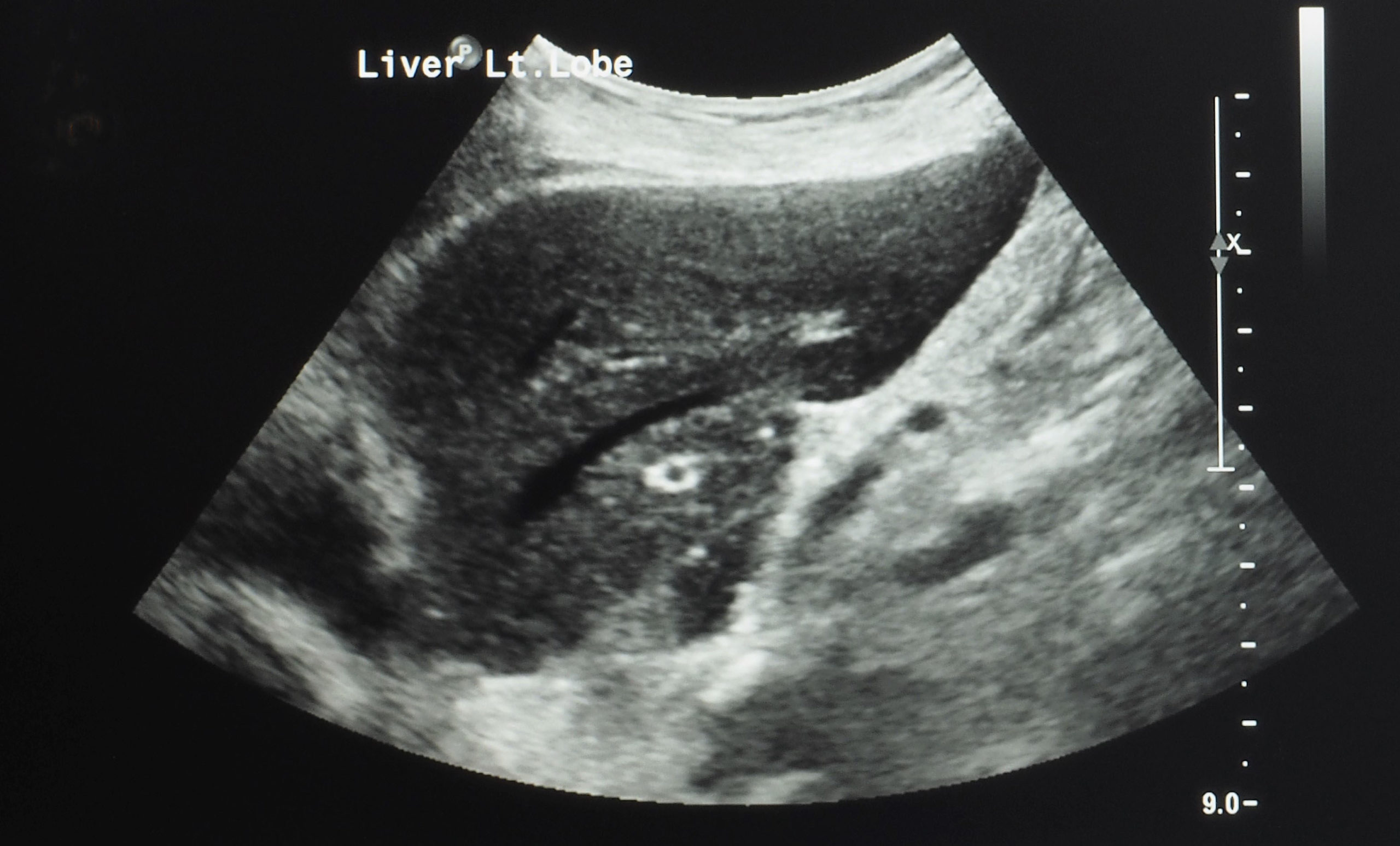
Liver and kidney ultrasound image
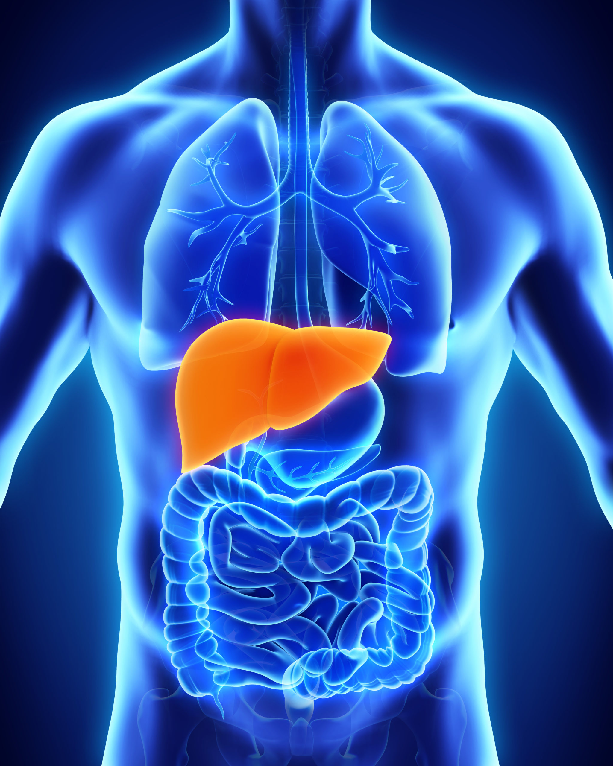
Illustration of the liver
Pelvic
Pelvic ultrasound includes transabdominal views of the female organs including the uterus, endometrium, cervix, vagina, ovaries (if not obscured by bowel), adnexas, and fallopian tubes. It is used to detect and/or monitor endometrial thickening, uterine fibroids or scars, ovarian cysts and masses, and tumors. If additional views are needed, then a transvaginal exam may be performed.
This may be valuable for:
• Pelvic pain
• Abnormal bleeding
• Infertility
• Fibroids
Ultrasound Resources & Articles:
Pelvic transabdominal exam
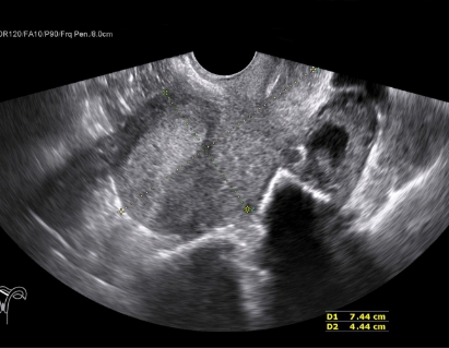
Uterus image
Female reproductive system
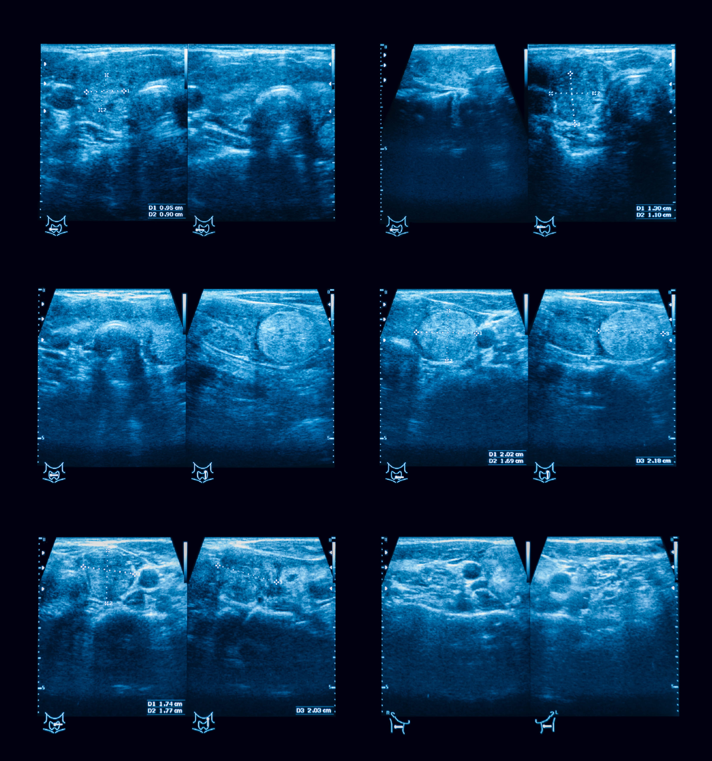
Renal (Kidneys & Bladder)
A renal ultrasound examines your right and left kidney, as well as your bladder to evaluate the size, detect cysts, tumors, obstructions, hydronephrosis, fluid collections, kidney stones, blockage of urine flow, and possible infections. The kidneys are the filtration system of your body. They filter the waste products out of your blood. The waste products then leave your body as urine.
A “post void” is also done with this exam. This requires you to come to the test with a full bladder so that the sonographer can get a volume of your bladder before and after you empty it.
This may be valuable for:
• Kidney stones
• Frequent urination
• Urinary tract infections
• Diabetes
Ultrasound Resources & Articles:
Renal ultrasound exam
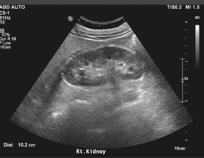
Ultrasound image of a kidney
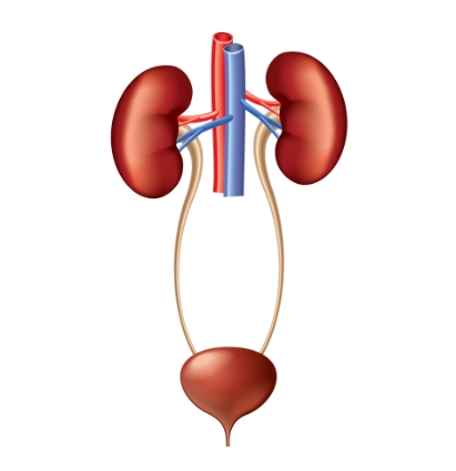
Illustration of the urinary system
Thyroid
The thyroid gland is located in front of the neck just above the collar bones and is shaped like a butterfly. The thyroid gland makes the thyroid hormone, which helps to regulate a variety of body functions including how fast the heart beats, regulates metabolism, controls how quickly the body uses energy, makes proteins, and also controls how sensitive the body is to other hormones.
It is very common for patchy areas or nodules to develop in the thyroid that may or may not be felt on the skin surface. Ultrasound is very sensitive and shows many nodules that cannot be felt. The vast majority of these are benign regions of thyroid tissue that pose no health risk. The minority of these are true tumors of the thyroid and may require further diagnosis, biopsy, or treatment. We examine the thyroid for size, lumps, nodules, or masses. It is also used to monitor existing growths for stability or change.
This may be valuable for:
• Thyroid nodules or lumps
• Abnormal thyroid function test
• Difficulty swallowing
• Hypo/Hyperthyroidism
Thyroid Resources:

Thyroid exam
Ultrasound image of a thyroid nodules
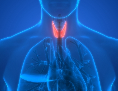
Illustration of the thyroid gland
Here’s how it works.
Explore ultrasound pricing and packages.
Breasts & Underarms
$295
(40 minutes)
Examines both breasts and underarm lymphatic glands. Also used to determine if any lumps are solid masses or fluid-filled cysts.
Women’s Health Check
$725
(80 minutes)
Includes breasts and pelvic organs (trans abdominal views), then choose between right upper abdomen or thyroid.
Complete Abdomen
$265
(40 minutes)
Includes liver, pancreas, gallbladder, spleen, right and left kidney.
Pelvic
$255
(40 minutes)
Abdominal views of the female reproductive organs. Please note, factors such as client preparation and body type may determine the image quality of the organs.
Breasts & Complete Abdomen
$525
(80 minutes)
Includes both breasts, lymph nodes, liver, pancreas, gallbladder, spleen, right and left kidney.
Thyroid
$220
(30 minutes)
Examines the thyroid to assess size, lumps, nodules, or masses.
Breast Thermography & Ultrasound Combo
$499
(60-80 minutes)
(Reg. $390) Includes your initial breast thermography & ultrasound appointment.
Women’s Health Check Thermography & Breast Ultrasound
$649
(80 minutes)
Includes thermography of both breasts, abdomen, thyroid, carotids, head & neck and bilateral breast ultrasound.
Full Body Thermography & Breast Ultrasound
$739
(1 hr 40 minutes)
Includes a full body thermography and bilateral breast ultrasound.
Thermography & Ultrasound Women’s Health Check
$1069
(2 hours 40 minutes)
Includes both women’s health check screenings. Thermography includes both breasts, abdomen, thyroid, carotids, head & neck. Ultrasound includes breasts and pelvic organs, then choose between right upper abdomen or thyroid.
Frequently Asked Questions
Can breast ultrasound detect cancer?
A breast ultrasound may increase your chances of detecting breast cancer from approximately 48% to 97% than with a mammogram alone, especially for women with dense breasts. It can detect structural masses that may indicate tumors or suspicious cysts which may require a biopsy for a diagnosis. Please note that no breast screening can diagnose cancer. Only a biopsy provides an actual diagnosis.
I have dense breast tissue, would ultrasound be helpful for me?
Breast ultrasound is highly beneficial for women with dense breast tissue because there are more ducts and glands, more fibrous tissue, and less fatty tissue. Traditional testing is less sensitive to these characteristics. For instance, mammograms may not be able to differentiate between breast cancer and dense tissue since they both may appear white on the image.
Are breast ultrasound results immediate and who interprets my images?
Breast ultrasound results are immediately sent to a radiologist for a review then the completed report is emailed directly to you within two weeks. If you need your results ASAP, there is a rush option available for an additional fee. Our technicians do not discuss findings during the appointment but, once the radiologist completes the report we are happy to answer any questions you may have.
Is breast ultrasound better than a mammogram?
These are two different types of tests—one does not replace the other and both are breast screenings that can detect cancer or abnormalities. Mammograms use radiation and compression while ultrasounds use sound waves.
A breast ultrasound may increase your chances of detecting breast cancer from approximately 48% to 97% than mammograms alone, especially for women with dense breasts because there is more fibrous tissue and less fatty tissue. Traditional testing is less sensitive to these characteristics. For instance, mammograms may not be able to differentiate between breast cancer and dense tissue since they both may appear white on the image.
Is breast ultrasound safe?
Yes. A breast ultrasound is safe because it uses sound waves (or echoes), rather than radiation, to visualize the structures of your body. This means it’s safe even for pregnant women.
What can ultrasound detect?
It can identify and measure areas of concern such as suspicious masses, tumors, cysts, blood clots, and blockages in areas such as breast tissue, lymph glands, major organs, and the vascular system. Ultrasound can also be used in a proactive manner to gain insight into your health.
Where can I get an ultrasound scan near me?
You can schedule an ultrasound screening at one of our six convenient ERLY Wellness locations in the Greater Atlanta area. Explore our locations to find an office near you.
What if my ultrasound results show a suspicious finding?
The Radiologist will make follow-up recommendations that may range from another ultrasound in six months to monitor any changes to additional testing and/or visiting your physician.
How often should I get a breast ultrasound?
If there are no abnormal findings, then many women will schedule annual screenings to monitor their breast health.
Do pregnancy, breastfeeding, or implants interfere with ultrasound?
No. Ultrasound is safe and effective for all of the above.